Esophagus Ct Scan Images - Dysphagia Lusoria presenting with Pill-induced ... / During an endoscopy, a biopsy may be taken.. These include physical examination and imaging tests such as ct scan, pet scan, and endoscopy. Ct was developed independently by a british engineer named sir godfrey hounsfield and dr. Early symptoms include acid reflux and difficulty swallowing, but symptoms are often not obvious until a later stage. This drink may taste unpleasant. They're carried out in hospital by specially trained operators called radiographers, and can be.
Confirmation or exclusion of malignant tumors; This provides a series of images from many different angles. Imaging tests for esophagus cancer. Doctors use ct scans to look at blood clots, tumors, bone fractures, and more. The esophagus may also be imaged using a flexible camera inserted into the esophagus, in a procedure called an endoscopy.
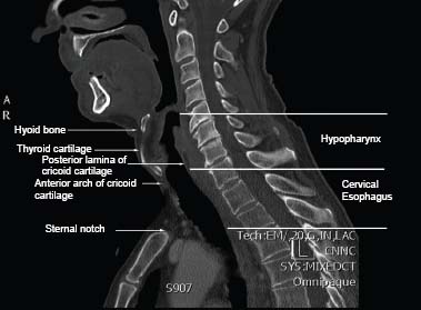
Hypopharynx and Cervical Esophagus Cancer | Ento Key from entokey.com Mid esophageal strictures and ulcers are suspicious for barrett's esophagus. This drink may taste unpleasant. On the left are ct images of a patient with large uphill varices secondary to cirrhosis with portal hypertension. These include physical examination and imaging tests such as ct scan, pet scan, and endoscopy. Esophageal cancer, ct scan with contrast, coronal image. The relative sensitivity of fluoroscopy, radionuclide scans. The esophagus is the tube that carries food and liquid to your stomach. Even in patients with symptoms consistent with esophageal spasm, thickening seen on ct scan images should not be dismissed as muscular hypertrophy secondary to the esophageal spasms without.
Imaging tests for esophagus cancer.
Ct scans show the anatomy quite well (compare normal ct scans here with typical esophagus cancer here and other pictures below). Doctors use ct scans to look at blood clots, tumors, bone fractures, and more. The esophagus may also be imaged using a flexible camera inserted into the esophagus, in a procedure called an endoscopy. This technique is used to prevent passive regurgitation during the induction of anesthesia in patients at high risk for regurgitation. Teaching files with ct medical imaging and case studies on anatomical regions including adrenal, colon, cardiac, stomach, pediatric, spleen, vascular, kidney, small bowel, liver, chest | ctisus. The esophagus is the tube that carries food and liquid to your stomach. Ct scans are also referred to as computerized axial tomography. Esophageal cancer (cancer of the esophagus). Ct imaging can also show wall thickness and evaluate for mediastinal involvement/general extent of disease beyond the mucosa. Treatment includes surgery, chemotherapy, and radiotherapy. Williford et al (38) reported. The analysis of pet/ct scans is complicated and requires a radiologist with experience in both pet/ct and head and neck imaging. The relative sensitivity of fluoroscopy, radionuclide scans.
Diagnostic medical test ct scan of the esophagus including diseases and symptoms diagnosed or ruled out by this test. Ct scans are also referred to as computerized axial tomography. Cricoid pressure occludes the esophagus (e) by compressing it between the cricoid cartilage (cc) and the body of the sixth cervical vertebra (c6). Esophagogastroduodenoscopy is more sensitive than any other method in the detection of esophageal stricture as well as esophagitis; • the accuracy of staging by esophagography, esophagoscopy, endoscopic ultrasonography, and ct scan for t staging was 80% and for n staging 72% with a sensitivity of 78.
Esophagus- Leiomyoma distal esophagus, coronal CT - YouTube from i.ytimg.com Manometry is considered the reference standard for evaluation of esophageal dysmotility. Esophageal cancer is a malignant tumor of the esophagus, the tube that connects the throat with the stomach. This provides a series of images from many different angles. The esophagus may also be imaged using a flexible camera inserted into the esophagus, in a procedure called an endoscopy. The esophagus is the tube that carries food and liquid to your stomach. Like one piece in a loaf of bread. Confirmation or exclusion of malignant tumors; Ct scans are also referred to as computerized axial tomography.
The esophagus is the connecting tube between the pharynx and stomach that functions to transport ingesta and fluids.
Ct was developed independently by a british engineer named sir godfrey hounsfield and dr. Imaging tests for esophagus cancer. Esophagus and gastroesophageal ct scanning. This technique is used to prevent passive regurgitation during the induction of anesthesia in patients at high risk for regurgitation. If you would like a large, unwatermarked image for your web page or blog, please purchase the appropriate license. An abdominal ct scan is an imaging method. Diagnostic medical test ct scan of the esophagus including diseases and symptoms diagnosed or ruled out by this test. Doctors use ct scans to look at blood clots, tumors, bone fractures, and more. Manometry is considered the reference standard for evaluation of esophageal dysmotility. Mid esophageal strictures and ulcers are suspicious for barrett's esophagus. The relative sensitivity of fluoroscopy, radionuclide scans. The analysis of pet/ct scans is complicated and requires a radiologist with experience in both pet/ct and head and neck imaging. Axial ct scan of trachea and esophagus.
Treatment includes surgery, chemotherapy, and radiotherapy. Everything you need to know about computed tomography (ct) & ct scanning. You may not embed one of our images on your web page without a link back to our site. Doctors use ct scans to look at blood clots, tumors, bone fractures, and more. Confirmation or exclusion of malignant tumors;
Anatomy of the Esophagus from www.aboutcancer.com Even in patients with symptoms consistent with esophageal spasm, thickening seen on ct scan images should not be dismissed as muscular hypertrophy secondary to the esophageal spasms without. Ct scans are sometimes referred to as cat scans or computed tomography scans. An abdominal ct scan is an imaging method. The esophagus is the connecting tube between the pharynx and stomach that functions to transport ingesta and fluids. Esophageal cancer forms inside the esophagus — a hollow, muscular tube about 10 inches long that carries food and drink from the mouth to the stomach. Cricoid pressure occludes the esophagus (e) by compressing it between the cricoid cartilage (cc) and the body of the sixth cervical vertebra (c6). Esophagi or esophaguses) is a muscular tube that conveys food and the cervical esophagus begins at the upper esophageal sphincter, which is formed by the cricopharyngeus muscle 6. Confirmation or exclusion of malignant tumors;
Early symptoms include acid reflux and difficulty swallowing, but symptoms are often not obvious until a later stage.
Ct imaging can also show wall thickness and evaluate for mediastinal involvement/general extent of disease beyond the mucosa. This drink may taste unpleasant. The esophagus is the tube that carries food and liquid to your stomach. You may not embed one of our images on your web page without a link back to our site. Teaching files with ct medical imaging and case studies on anatomical regions including adrenal, colon, cardiac, stomach, pediatric, spleen, vascular, kidney, small bowel, liver, chest | ctisus. Everything you need to know about computed tomography (ct) & ct scanning. These include physical examination and imaging tests such as ct scan, pet scan, and endoscopy. During an endoscopy, a biopsy may be taken. Williford et al (38) reported. Esophageal cancer (cancer of the esophagus). 36 the cervical portion of the esophagus begins dorsal to the caudal border of the cricoid cartilage, inclines to the left of the trachea as it runs caudally, and ends at the thoracic inlet. Imaging tests for esophagus cancer. Ct scans are sometimes referred to as cat scans or computed tomography scans.


Belum ada Komentar untuk "Esophagus Ct Scan Images - Dysphagia Lusoria presenting with Pill-induced ... / During an endoscopy, a biopsy may be taken."
Posting Komentar